
Seeing the invisible injury
Using neuro-visual experts to prove mild traumatic brain injury
When it comes to Mild Traumatic Brain Injury (mTBI) cases, the defense playbook is almost always the same: No objective evidence. CT/MRI normal. Symptoms are subjective.
It’s effective but untrue. It’s effective because it is built on a half-truth: standard CT and conventional MRI often, more than 90% of the time, miss mild TBI. It’s untrue because there is objective evidence available to us, if we are willing to look. Specifically, we need to take a closer look at the visual system.
Thirty to fifty percent of the brain is devoted to seeing. When the brain is injured, the seeing breaks down in ways that produce numbers, charts, prescriptions, and follow-up appointments – not just patient complaints. Our job is to find those numbers and put them in front of a jury.
Why mTBI is so hard to prove
The definition
The World Health Organization Collaborating Task Force and the Centers for Disease Control define mTBI as an acute brain injury resulting from mechanical energy to the head from external physical forces. Operational criteria include one or more of the following: confusion, disorientation, or loss of consciousness for 30 minutes or less; post-traumatic amnesia of less than 24 hours; or transient neurological abnormalities such as focal signs, seizure, or intracranial lesions not requiring surgery – with a Glasgow Coma Scale of 13 to 15 at 30 minutes post-injury or later.
The peer-reviewed literature is in consensus that people with mTBI have a high degree of disruption of both visual input to the brain (the afferent visual system) and the brain’s control of ocular motor systems (the efferent visual system), with both objective and subjective findings. A careful neuro-ophthalmic assessment is the road map; for a detailed treatment of these systems, see the Trial Guides volume Brain Injuries: A Multidisciplinary, Illustrated Guide.
The Defense Health Agency (DHA) has framed the term Ocular Motor Rehabilitation (OMR) for Ocular Motor Dysfunction (OMD) as a deficit to Ocular Motor Function (OMF), to address symptoms such as blurry vision, double vision, tracking issues, headaches, dizziness, photosensitivity, and imbalance in patients who have suffered mTBI/concussion, within the Department of War (DOW) and Veterans Affairs (VA) systems of care, for the purpose of developing shared interprofessional definitions. OMDs include dysfunction of ocular alignment (tropias and phorias), movement (pursuit and saccade), binocular coordination (version and vergence), and accommodation. They impact balance, spatial orientation, proprioception, and visual-motor coordination – even when the eyes themselves are structurally normal.
The classic symptoms
The classic mTBI symptom list is familiar: headache, brain fog, irritability, photophobia, fatigue, sleep disruption, slowed processing, memory problems, cognitive impairment. Every one of them is patient-reported. None of them shows up on a scan. That is the whole architecture of the defense.
The defense script
We all hear different variations on the same theme: “self-reported,” “symptom magnification,” “litigation-driven,” “Grade 1 concussion,” “should have resolved in seven to ten days,” “no objective basis for ongoing complaints.” These are not effective. The defense is banking on the fact that when most people imagine “brain injury,” they picture a coma, a brain bleed, etc. They do not picture a software engineering manager who has difficulty scrolling a screen or a surgical ophthalmologist who needs prism glasses and can no longer perform surgery. Our job, along with our experts, is to teach the jurors that brain injury is much broader than common misconceptions.
CACI 3903 allows recovery for the loss of “physical, mental, and emotional well-being” once we prove causation. Causation is where these cases live or die. Neuro-visual evidence can help prove causation in brain injury cases, including mild traumatic brain injury.
mTBI is a process, not an event
The defense wants the jury to believe an mTBI is a single moment that resolves in days, maybe weeks. However, brain injury is a process. That process is called a neurological cascade.
The primary injury is mechanical – acceleration, deceleration, rotation. When the brain, suspended in fluid inside a hard skull, moves, it strikes the inside of the skull at the point of impact (coup), then rebounds and strikes the opposite side (contrecoup). Rotational and shear forces stretch and tear axons – the brain’s wiring – at the microscopic level. This is diffuse axonal injury, which cannot be detected by standard CT and conventional MRI.
The secondary injury unfolds over hours, days, and weeks: glutamate excitotoxicity, calcium influx, mitochondrial failure, oxidative stress, neuroinflammation. The neurons that survived the first hit are now sitting in a hostile environment. Some recover; some don’t. The defense narrative of “seven to ten days and gone” assumes a degree of cellular forgiveness the brain does not actually have.
The “mild” in mild traumatic brain injury is a Glasgow Coma Scale category, not a measure of how the injury impacts a person’s life. Mild means the injury hides better, not that the injury is small.
Why the visual system is uniquely vulnerable
Vision is not something the eyes do. It’s something the brain does. The eyes are hardware. The brain is software. Changes in the brain’s connectivity or function are routinely expressed through the eye’s machinery, which is why the visual system is uniquely positioned as an objective biomarker for brain dysfunction.
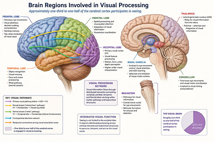
Thirty to fifty percent of the cerebral cortex is involved in visual processing: primary visual cortex (occipital lobe), parietal and temporal lobes, frontal eye fields in the frontal cortex, the brainstem, thalamus, basal ganglia, cranial nerves, cerebellum, and the visual tracts). Because mTBI generates rotational acceleration forces that stretch the brain’s long white-matter tracts, the visual pathway – optic nerve, optic tract, optic radiations, superior longitudinal fasciculus, inferior fronto-occipital fasciculus, etc. – is a natural target.
Walk through the visual pathway
Light enters the eye through the pupil and strikes the retina (an extension of the central nervous system), where photoreceptors convert light into neural signals. Those signals travel through the optic nerve (CN II) toward the brain. At the optic chiasm, nasal fibers from each retina cross to the opposite side; temporal fibers stay on the same side. The combined fibers continue as the optic tracts. Most synapse in the lateral geniculate nucleus of the thalamus, where input is organized and relayed to the visual cortex in the occipital lobe for processing of form, color, and motion. Some fibers diverge to the Edinger-Westphal nuclei in the midbrain, adjacent to CN III, controlling pupillary constriction and accommodation.
From the occipital lobe, higher-order processing proceeds to the parietal lobe for spatial orientation, the frontal lobe for ocular motor response, and the temporal lobe for object recognition. Supranuclear pathways – brainstem, cerebellum, basal ganglia, and cortex – generate saccadic and smooth-pursuit eye movements; the cerebellum fine-tunes them. Infranuclear pathways from the CN III, IV, and VI nuclei in the midbrain and pons control the extraocular muscles. The medial longitudinal fasciculus coordinates CN III and CN VI activity and, together with the infranuclear pathways, produces the vestibulo-ocular reflex (VOR).
Neuro-visual experts
Neuro-ophthalmologists are medical doctors (M.D.) who diagnose and treat neurological diseases with ophthalmic manifestations. They are trained in medicine, general ophthalmology, and neurology, with a focus on the brain disease rather than the eye itself. Neuro-optometrists hold a doctorate in vision science (O.D.). They are experts in ocular motor function, primary eye care, and the medical, non-surgical, management of ocular disease.
Both produce incontrovertible evidence including dated, repeated, numerically scored examinations performed in the ordinary course of clinical care, often beginning long before any lawyer is involved.
The neuro-visual exam – What gets measured objectively
A neuro-ophthalmic exam evaluates the afferent and efferent visual pathways. Findings include both objective measurements and patient-reported symptoms. When the two corroborate one another – and corroborate the clinical history, the clinical exam, and the neuroimaging – the diagnosis of mTBI is established to a reasonable degree of medical probability. It is a multidisciplinary process: neurologists, optometrists, neuro-ophthalmologists, occupational and physical therapists, audiologists, speech pathologists, and neuropsychologists each opine, and the diagnosis is built from the convergence of their findings.
Objective findings
- Pupillary findings: dilated or poorly reactive pupils; afferent pupillary defect (APD).
- Optic nerve pallor and traumatic optic neuropathy on optical coherence tomography (OCT) and posterior segment exam.
- Strabismus (tropia) and phorias documented on cover/uncover, Hirschberg corneal light reflex, and version testing in nine cardinal positions of gaze.
- Cranial nerve III, IV, or VI palsies and paresis, with measured prism diopters of misalignment.
- Saccadic intrusions, deficient pursuit, reduced positive fusional vergences, abnormal near point of convergence (NPC).
- Visual field defects on threshold perimetry, often localizing the lesion within the brain.
Photosensitivity
Photosensitivity is common in mTBI and remains largely subjective on direct testing – the workup is to rule out anterior and posterior segment causes (dry eye, meibomian gland dysfunction, neurotrophic keratitis, conjunctivochalasis, iris atrophy, retinal pathology). The literature implicates central sensitization of the trigeminothalamic pathway. The trigeminal nerve (CN V) communicates with the thalamus, the brain’s sensory gatekeeper. When the thalamus is under assault in mTBI, it becomes hyperexcitable. Normal light becomes overwhelming. The patient who walks into the deposition wearing sunglasses indoors is not performing for the camera. The trigeminothalamic pathway is doing its job badly.
Every meaningful test in a neuro-visual workup produces a number or an image. Near point of convergence is centimeters. Stereopsis is seconds of arc. Phorias and tropias are prism diopters. Visual fields and OCTs are printed images.
This is the antidote to the “it’s all subjective” defense.
Suplizio v. Proliance Surgeons
Jason Suplizio was a 50-year-old senior software engineering manager at Adobe, a mountain biker, master’s-degreed, and a qualified pilot candidate. On June 24, 2021, he was dropped on his head while under general anesthesia for a routine shoulder surgery. The CT and MRI were normal. The defense argued that Mr. Suplizio’s complaints were ‘entirely subjective.’
We were able to show the chart of his treating optometrist, Dr. Lisa Dok, F.A.A.O. Over more than two years, Dr. Dok saw Jason 61 times. She measured his near point of convergence and watched it move. She measured stereopsis and watched it move. She prescribed prism glasses and vision therapy. She paused therapy when she suspected an auditory processing component was holding back his visual recovery, referred him out, and resumed treatment. Most of this was objective evidence of Mr. Suplizio’s brain injury.
Dr. Dok told the jury that approximately 80% of concussion patients develop convergence insufficiency. She told them Jason’s findings were “more along the lines of someone who has post-concussion syndrome with visual findings, rather than just someone who just has a pure convergence insufficiency.” She told them his injuries were permanent.
The defense argued pre-existing convergence insufficiency from age 16. Dr. Dok answered with a 33-year asymptomatic interval, during which Jason passed a pilot medical, earned a master’s degree, and read 20 books a year. She told the jury, based on objective medical evidence, that the fall created a new injury.
Tong v. American Auto Auction Group
Dr. Ly Ti Tong was a practicing surgical ophthalmologist. In April 2021 she was rear-ended. The CT was normal. The conventional MRI was normal.
After the collision, for the first time in her life, Dr. Tong needed a prism prescription. Her treating optometrist examined her in March 2022 and wrote the prism.
The defense neurologist was hired to tell the jury that his neurological exam was ‘normal.’ We were able to show through cross-exam that the general neurologist’s acute neurological exam is not designed to find convergence insufficiency, saccadic intrusions, or accommodative dysfunction.
Dr. Samuels: A pilot who cannot fly
A 49-year-old commercial airline pilot was involved in a rear-end motor vehicle collision. He hit his head against the window on the left side, with possible loss of consciousness, and was diagnosed in the emergency department with mTBI and orthopedic injuries. He then developed accommodative insufficiency (worse in the left eye), convergence insufficiency, intermittent strabismus, deficient saccadic eye movements, and diplopia. His ocular motor system was under siege. He was no longer fit for duty. Federal Aviation Administration standards automatically remove pilots with ocular motor dysfunction from the cockpit. He lost his career, which was devastating both financially and emotionally. He will likely never return to aviation. The case is ongoing.
Cross-examining the defense neurologist with neuro-visual evidence
- Establish the limits of the screening exam. A routine cranial nerve check tests gross function. It is not a substitute for a neuro-ophthalmic evaluation, an OCT, a threshold visual field, or measured prism cover testing.
- Establish what the witness did not review. Most defense neurologists have not read the treating optometrist’s records.
- Establish what those records show. Numbers. Dates. Diagnoses. Prescriptions. Get the witness to agree that none of this is patient self-report.
- Close the loop on causation. Establish that the patient had no pre-injury visual symptoms and that the post-injury records are objective. This proves that something between point A and point B caused the change. The trauma is more likely that cause.
- The ‘invisible injury’ is a defense frame. Stop accepting it.
Brain injury is not invisible – it’s connected to one of its most measurable functions: the visual system. When we build our cases on charts, visits, measurable convergence, a prism prescription, a visual field printout, etc., the defense’s ‘subjective’ and ‘invisible’ themes fall flat.
Of course, we don’t stop there. Brain injury is visible in the people it affects, which includes the plaintiff, of course, but also includes the people around them: their family, co-workers, friends, neighbors. Lay witnesses make brain injury visible and visceral and are powerful evidence in the courtroom.
Practical tips
- Retain a neuro-visual expert early.
- Use the treating chart as an exhibit.
- Make a measurements timeline
- Take treaters’ depositions for use at trial and emphasize the patient physician relationship.
- Cross the defense neurologist with the treating optometrist’s records.
- Gather lay witnesses who have direct experience with the ‘before’ and ‘after.’
Courtney Rowley is a national trial lawyer, published author, and mother. She represents clients in catastrophic injury, medical malpractice, toxic tort, and wrongful death cases. She is CAALA Trial Lawyer of the Year 2025. She and her husband, Nick, have spent their careers trying cases together as equal partners, and have been politically active in defending the civil justice system – helping reform California’s MICRA caps on medical malpractice damages and, this year, fighting to protect civil rights against the Uber ballot initiative. She has taught trial skills across the country for more than 15 years, including several years teaching at Gerry Spence’s Trial Lawyers College.
Marianna Samuels, O.D., is a board-certified optometrist and clinical researcher based in San Diego. A Diplomate of the American Board of Optometry, she practices medical optometry at North County Eye Center, where she also serves as Medical Director of the Refractive Surgery Clinic. Dr. Samuels earned her Doctor of Optometry from the University of California, Berkeley, School of Optometry, graduating magna cum laude. Dr. Samuels is a recognized authority on traumatic brain injury and its effects on the visual system. She authored the chapter on neuro-ophthalmologic assessment in TBI for Brain Injuries: A Multidisciplinary, Illustrated Guide, and she serves as an expert witness on TBI-related eye and visual-pathway injuries, primarily for plaintiffs.
Courtney Rowley
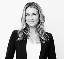
Courtney Rowley is a national trial lawyer, published author, and mother. She represents only people – never corporations, insurers, or institutions – in catastrophic injury, medical malpractice, toxic tort, and wrongful death cases. She is CAALA Trial Lawyer of the Year 2025.
Most recently, Courtney co-led the civil prosecution of Monsanto on behalf of more than 150 teachers, students, and parents poisoned by PCBs at the Sky Valley Education Center in Monroe, Washington. After three years of litigation and multiple trials, Monsanto settled the entire inventory of pending Sky Valley cases.
Courtney's work extends beyond the courtroom. She and her husband, Nick, have spent their careers trying cases together as equal partners, and have been politically active in defending the civil justice system – helping reform California's MICRA caps on medical malpractice damages and, this year, fighting to protect civil rights against the Uber ballot initiative.
Closest to her is Trial By Woman, the nonprofit she co-founded with Theresa Hatch. What began as a small effort to teach and support women trial lawyers has grown into a national community of powerful attorneys.
Courtney has authored four books: Trial By Woman; Voir Dire and Opening Statement; Running with the Bulls: How to Win Top Dollar Settlements; and Damages Evolving, with David Ball and Artemis Malekpour. She has taught trial skills across the country for more than 15 years, including several years teaching at Gerry Spence's Trial Lawyers College.
She grew up in Oxnard, California, and studied at USC and Loyola Law School. She and Nick live in Montana with their children.
Marianna Samuels

Marianna Samuels, O.D., is a board-certified optometrist and clinical researcher based in San Diego. A Diplomate of the American Board of Optometry, she practices medical optometry at North County Eye Center, where she also serves as Medical Director of the Refractive Surgery Clinic. Dr. Samuels earned her Doctor of Optometry from the University of California, Berkeley, School of Optometry, graduating magna cum laude. Dr. Samuels is a recognized authority on traumatic brain injury and its effects on the visual system. She authored the chapter on neuro-ophthalmologic assessment in TBI for Brain Injuries: A Multidisciplinary, Illustrated Guide, and she serves as an expert witness on TBI-related eye and visual-pathway injuries, primarily for plaintiffs.
Copyright ©
2026
by the author.
For reprint permission, contact the publisher: Advocate Magazine